 Up
Up
|
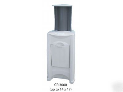
The Navigator requires only minimum computer knowledge for operation. All systems are installed and in-serviced by factory trained personnel and come with unlimited life time toll-free technical support. The NAVIGATOR is ideally suited for both small and large facilities and is compatible with multi-room or mobile application. Intra-oral, upper and lower dental arcade x-ray capability for small and large animal applications is standard. * Elimination of film processing * No odors or hazardous waste material * Faster, almost immediate real time image display * Decrease of patient x-ray dosage * Image manipulation for an enhanced diagnosis (zoom, contrast, tilt, color, special filters, archiving, etc.) * Less expensive to operate (eliminate chemicals, film and processors * Less time consuming- Little maintenance, no processor cleaning * Better reliability (consistent & repetitive high quality images) * Less patient stress, increased image diagnostic information & manipulation with less patient movement * Capable of transmitting images via email * Images are stored in the computer for further reference, easy accessibility and transfer * Archival, long-term storage and retrieval of images Items included with Digital NAVIGATOR: * High powered computer desktop or laptop system with cables and accessories * 21" high contrast standalone LCD monitor provided with desktop system * Networkable control and scanning VetRay software * Image processing software allowing image manipulation, storage/transfer * High-resolution printer with color and grayscale printing *Technique charts for small and/or large animal The Navigator is designed to replace all your x-ray films with the highest quality digital images. Digital Radiology eliminates the inconvenience and high costs associated with the use of film technology. The Digital Navigator System uses cassettes in a similar manner to those used in conventional film-screen radiology. These cassettes, however, do not contain film, but an advanced photostimulable phosphor plate. After exposure, the plate is removed from the cassette with out the use of a darkroom, placed in the CR reader, where a laser light stimulates the particles in the plate to luminesce. The light is collected using a light guide and a high resolution photo multiplier tube where it is converted to a digital signal. The image can then be, viewed, enhanced, stored, transferred or printed via a laptop or dedicated computer work station. Once the plate has been scanned it is erased by exposure to a bright white light, which prepares it for reuse. By listing in this category, you certify that you will comply with all such laws. In addition, you must include: 1) your complete business name 2) city and state of the business and 3) type of business (i.e., hospital, medical office, manufacturer, distributor, broker, etc.) If you have questions about legal obligations regarding sales of medical devices, you should consult with the FDA's Center for Devices and Radiological Health: Links: http://www.fda.gov/cdrh/devadvice/ or http://www.fda.gov/cdrh/industry/support/index.html |
